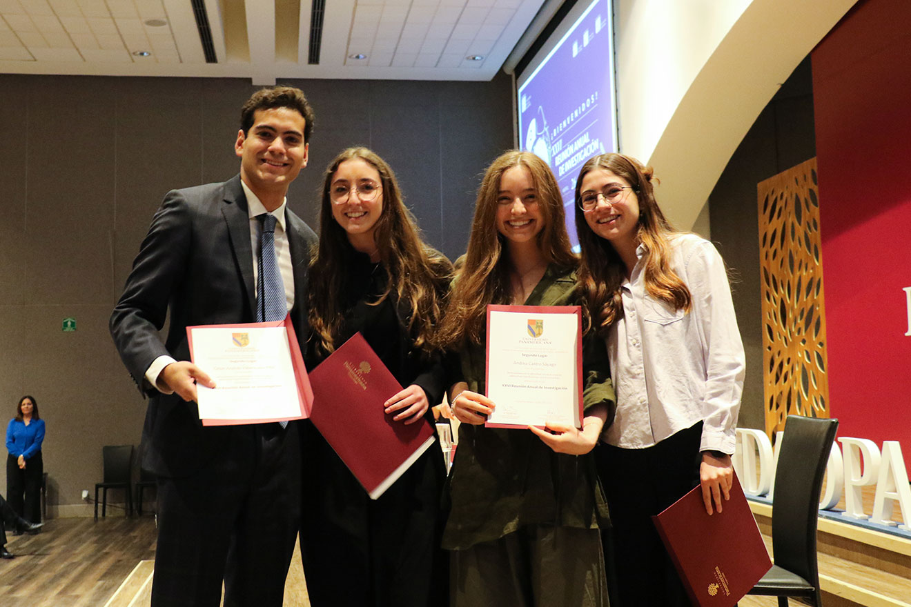
Aguascalientes, Ags., September 19, 2024.- Ricardo Espinosa, research professor at the School of Engineering of our Panamerican campus in Aguascalientes, Aguascalientes, Mexico, has been a member of our Panamericana campus Aguascalientes shares with us in detail about his most recent research "Color-aware Exposure Correction for Endoscopic Imaging using a Lightweight Vision Transformer", which he conducted together with students Javier Eluney Hernandez and Ulises Gallardo Rodriguez, with which they received the award for best student paper in one of the most important congresses in the area of medical imaging: the IEEE CBMS 2024.
A project for the benefit of health
"Saving lives" is the first phrase that Professor Ricardo Espinosa expressed when asked what was the main motivation behind this article. In Mexico, the fourth most common cancer is colon cancer, as about 15,000 new cases of this type of cancer are diagnosed each year, according to the Ministry of Health.
When analyzing the various problems that exist when performing an endoscopy, in this case focused on colonoscopy, it was mainly detected the limitation that a physician has when not spatially locating exactly where the endoscope is located, so there is the possibility of leaving important areas without inspecting or interesting findings.
The concern of being able to contribute to the problem led him to think about how it would be possible to insert Artificial Intelligence (AI) models that support better decision making by doctors, where through a specific algorithm health experts can observe details that had not previously been detected and thus lives can be saved, which is why he started this project.
One of the primary branches of this work is based on three-dimensional reconstruction of endoscopic images.
"Having a 3D reconstruction at hand, the physician will know exactly where the tip of the endoscope is inside the inspected organ and will also be able to find ulcers, bleeding or polyps more quickly and easily compared to a general endoscopy," he reports.


However, this is not the only problem. The illumination of the endoscope, being the only source of information, generates overexposed (very bright) or underexposed (very dark) images. In this sense, the idea was to generate a reliable 3D reconstruction that achieves stability in the illumination.
A unique and innovative algorithm
"What we did in this research was to generate an AI-based algorithm to be able to attack these problems of overexposure and underexposure that occur in endoscopic images to subsequently generate a three-dimensional shape of the inspected device in a more reliable way," he says.
Endo Vision 3D is a computer vision algorithm that reconstructs endoscopically inspected organs in 3D. It improves medical accuracy and efficiency by 30%, reduces surgical times by 20%, and allows lesions to be recorded and tracked over time.
One of the characteristics that make this algorithm unique and innovative is that it comprehensively attacks both variants of this problem: overexposure and underexposure, while other algorithms only attack one of the two variants.
To check the effectiveness of the algorithm, several interesting metrics are used such as -structuralsimilarity loss, PSNR- to measure how similar the image that the algorithm produced is to the original image, in this way the results can be compared against what was expected.
"The ultimate goal of this project is to help more doctors who suffer from these difficulties on a daily basis. If doctors had this system that in real time could be improving the illumination in each frame, simply with this solution they could have a better effectiveness in detecting diseases," he adds.